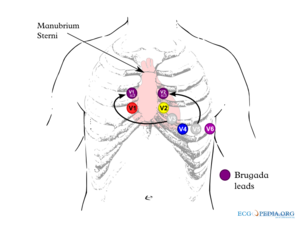
Brugada Type 2 Not Type 1 V1 and V2 may be placed in the 3 rd or even 2 nd intercostal spaces in order to elicit a type 1 Brugada pattern and is considered diagnostic. Demonstration and unmasking of the type 1 ECG pattern at the higher lead positions also have diagnostic value for Brugada syndrome similar to its.

Brugada Syndrome Brs This Picture Displays The Ecg Trace Of A Download Scientific Diagram
80 of Brugada syndrome diagnosed only after a cardiac arrest.

. However errors in placement of ECG leads can create artifacts mimic pathologies and hinder proper ECG interpretation. The diagnosis of the characteristic type-1 or coved type Brugada ECG figure 1 is made from the right precordial ECG leads see also Wilde 2002. Ago edited 1 yr.
12 To ensure consistent lead placement we incorporated a high precordial lead V1 and V2 at the 2nd intercostal space ECG into our. Please see the file description page for further information. 84 KB MIME type.
1 There is one true diagnostic of the Brugada pattern. And the caveats of improper lead positioning were recently reviewed. B Spontaneous type 1 Brugada ECG pattern in a 53-year-old man with aborted cardiac arrest implanted ICD and subsequently multiple appropriate shocks of the device.
Except some very rare palpitations doesnt have any other symptoms. This is the second of a two-part series discussing how to recognize and avoid these errors. A Fractionated QRS complex in a 25-year-old asymptomatic male patient with BrS ajmaline-induced type 1 Brugada ECG pattern.
Conclusion In our athletes without preexisting incomplete RBBB a Brugada-like pattern was easily obtained and highly prevalent with high placement of anterior precordial leads. Placement of the right precordial leads in a superior position up to the 2 nd intercostal spaces above normal can increase the sensitivity of the ECG for detecting the Brugada phenotype in some. It features large coved ST-segment elevations and T-wave inversions in leads V1V3.
A typical baseline Fig A and positive ECG Fig B are shown. The positivity was defined as inducible Type 1 Brugada pattern in atleast 2 right sided leads. In ECG 1 the base of the triangle horizontal PINK line formed by drawing lines from the peak of the r in lead V2 looks to be extremely close to 35 mm which is right at the upper limit qualifying for a Brugada-2 pattern.
Two others may suggest the disease. Proven VF or VT. It is characterised by a prominent coved.
Brugada Syndrome is an ECG abnormality with a high incidence of sudden death in patients with structurally normal hearts. Standard 12-lead ECG with V1V3 recorded from the fourth intercostal space and an additional. To assess the feasibility and reliability of using high precordial lead ECGs in conjunction with three new criteria for identifying true.
By contrast a type 2 Brugada pattern may often be found with these high leads are applied to healthy people especially in fit young males. Brugada Syndrome is a rare inherited cardiac arrhythmia syndrome that is characteristed by a coved-shaped atypical right bundle branchpattern on a 12-lead ECG Type-1 Brugada pattern ECG and is associated with ventricular arrhythmias and sudden cardiac death. Recently three criteria analyzing the ST segment and r width to identify true Brugada pattern ECGs have been described.
Brugada syndrome is a rare inherited arrhythmic disorder causing an increased risk of syncope and sudden death due to ventricular fibrillation. Brugada syndrome is diagnosed in patients with type 2 or type 3 ST-segment elevation in 1 lead among the right precordial leads V1 V2 positioned in the 2nd3rd or 4th intercostal space when a provocative drug test with intravenous administration of class I antiarrhythmic drugs induces a type I EKG morphology 3. A programmed electrical stimulation was performed on our patient and a sustained ventricular arrhythmia.
The use of precordial leads in the 2nd or 3rd intercostal space has been described in patients with suspected and known Brugada syndrome5 11. A higher right precordial lead placement than the conventional position at V1V3 can also be used to unmask the Brugada-type ECG in subjects without typical ECG changes or unequivocal signs. The Brugada syndrome may present with three different ECG patterns referred to as type 1 type 2 and type 2 Brugada syndrome ECG.
Placement of the right precordial leads in a superior position up to the second intercostal space above normal can increase the sensitivity of the ECG for detecting the Brugada phenotype both in the presence or absence of a drug challenge. Resting 12-lead ECG with standard precordial leads and ECG with precordial leads placed 1 Intercostal space above were performed after flecainide administration every 5 min for first 30 min and every 30 min thereafter until ECG became normal or upto 6 h. Diagnosis can only be considered appropriate with a positive ECG as well as at least one of the following.
Aims The authors sought to assess the value of the high right precordial leads RPL to detect the Type I Brugada ECG pattern in patients suspected of carrying Brugada syndrome BrS. Electrocardiography is a very useful diagnostic tool. An rSr pattern in leads V1-V2 can be observed when ECG leads are placed in the 2nd intercostal space.
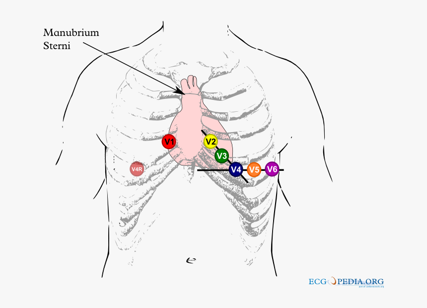
Three repolarisation patterns are associated with BS4 Figure 1 when found in more than one right precordial leads V1 to V3. Consider as cause of syncope in patients with family history of sudden death. Brugada_lead_placementpng 800 600 pixels file size.
Double check that lead placement is in the correct intercostal spaces and repeat ECG. Lead V1 from the 4th 3rd and 2nd ic. The mean age of sudden death is 41 with the age at diagnosis ranging from 2 days to 84 years.
Sensitivity of the ECG can be increased with alternative placement of ECG leads to the intercostal space above V1 and V2 figure 2. Brugada Syndrome is an abnormal ECG Right Bundle Branch Block Pattern with coved ST elevation over the right precordial leads of V1-V3 which leads to ventricular fibrillation VF and sudden cardiac death SCD in patients with structurally normal hearts. When gaining a 12-Lead ECG a configuration wherein the placements of V1 and V2 are positioned more cranially V1 ICS12 V2 ICS12 should be recorded as these leads can often give a clearer view of abnormal morphologies.
First described in 1992 by the Brugada brothers the disease has since had an exponential rise in the numbers of cases reported. It has been recognized as a clinical entity since 1992. 12-lead ECGs were recorded in a single male healthy subject in his mid.
The most typical and diagnostic is type 1 Brugada syndrome. Careful placement of leads is essential to avoid false positive findings. Correct recognition of the diagnostic Brugada syndrome ECG pattern.
Brugada Syndrome is reported to be responsible for 4 of all sudden deaths and 20. ECG recordings and even more with placement of precordial leads V1V2 and V3 in the 2 nd and 3 rd intercostal space which may bring out a typical Brugada pattern and should be routinely performed when the diagnosis is suspected but is uncertain on a standard ECG and in screening of family members of BS patients. Methods Ajmaline testing using 15-lead ECGs was performed in 183 patients suspected of carrying BrS.
Op 1 yr. Criteria for a Brugada-2 ECG pattern disappeared once the V1 and V2 electrodes were correctly positioned See ECG 2 in Figure-1. Imagepng This file is from a shared repository and may be used by other projects.
Male 19 years old. Does not meet criteria for a Brugada pattern could be an IRBB.
Brugada Syndrome Ecgpedia
Brugada Syndrome Ecg Placement Hd Png Download Kindpng
2

Positioning Of High Precordial Leads Hv1 To Hv6 For Procainamide Download Scientific Diagram

How To Perform And Interpret Provocative Testing For The Diagnosis Of Brugada Syndrome Long Qt Syndrome And Catecholaminergic Polymorphic Ventricular Tachycardia Circulation Arrhythmia And Electrophysiology
Ecg Interpretation In Brugada Syndrome Sciencedirect

Ecg Interpretation In Brugada Syndrome Sciencedirect

Raviele Antonio 在 Twitter 上 Figure Showing The Value Of High V1 V2 Leads Placement To Unmask Type 1 Brugada Ecg Pattern Dr Santangeli Abollmannmd Ecgtalk Adribaran Drfermingarcia Jorgeeromeromd Lluismont2 Manliomarquez Drjasonandrade
0 comments
Post a Comment